Basal Ganglia Clinical Anatomy Physiology Biology Diagrams
BlogBasal Ganglia Clinical Anatomy Physiology Biology Diagrams The basal ganglia approve or reject Trusted Source PubMed Central Highly respected database from the National Institutes of Health Go to source movement signals the brain sends, called modulating inhibition, enabling the use of specific muscles when taking action. The basal ganglia also receive sensory information from other parts of the body and use it to refine movement.
The basal ganglia are a subcortical collection of interacting clusters of cell bodies, and are involved in reward, emotional, and motor circuits. Functional anatomy of the basal ganglia. According to Steiner and Leite R. E. P., et al. (2009). Equal numbers of neuronal and nonneuronal cells make the human brain an isometrically scaled-up Tics are nonvoluntary movements of any part of the body that are rapid, sudden, repetitive but nonrhythmic, suppressible for at least a moment, and typically associated with an irresistible urge or premonitory sensation. et al. Evidence for segregated and integrative connectivity patterns in the human Basal Ganglia. The Journal of The basal nuclei (blue structures in the diagram) are essential in the control of movement, and regulation of mood and complex behaviors.. An older name for the basal nuclei is basal ganglia. However, we've made a big deal of telling you that a "ganglion" is a cluster of nerve cells outside the CNS, and the basal nuclei are definitely part of the CNS, so we'd be hypocritical and

Concise Medical Knowledge - Lecturio Biology Diagrams
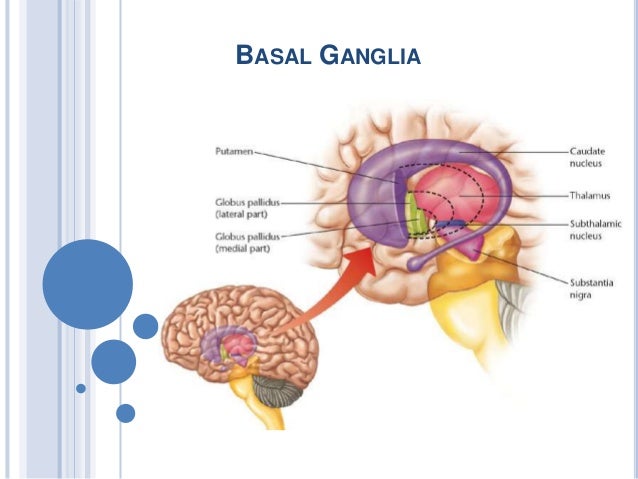
The basal ganglia consist of several large, anatomically distinct masses of gray matter situated in the core of the cerebral hemispheres among ascending and descending tracts of white matter and astride the brainstem. These constitute the striatum, comprised of the caudate and putamen, and the pallidum, comprised of the internal and external portions of the globus pallidus. The basal ganglia are a group of subcortical nuclei, meaning groups of neurons that lie below the cerebral cortex. The basal ganglia is comprised of the striatum, which consists of the caudate nucleus and the putamen, the globus pallidus, the subthalamic nucleus, and the substantia nigra The basal ganglia are primarily associated with motor control, since motor disorders, such as Parkinson's The basal ganglia are best known for how they help your brain control your body's movements. However, ongoing research continues to uncover other ways that the basal ganglia interact with other parts of your brain. Though experts continue to uncover more about the inner workings of the basal ganglia, there's much about them that remains

Basal ganglia are a group of subcortical nuclear agglomerations involved in movement, and are located deep to the cerebral hemispheres. Basal ganglia include the striatum (caudate nucleus Nucleus Within a eukaryotic cell, a membrane-limited body which contains chromosomes and one or more nucleoli (cell nucleolus). The nuclear membrane consists of a double unit-type membrane which is perforated
