Human Venous System Anatomy Biology Diagrams
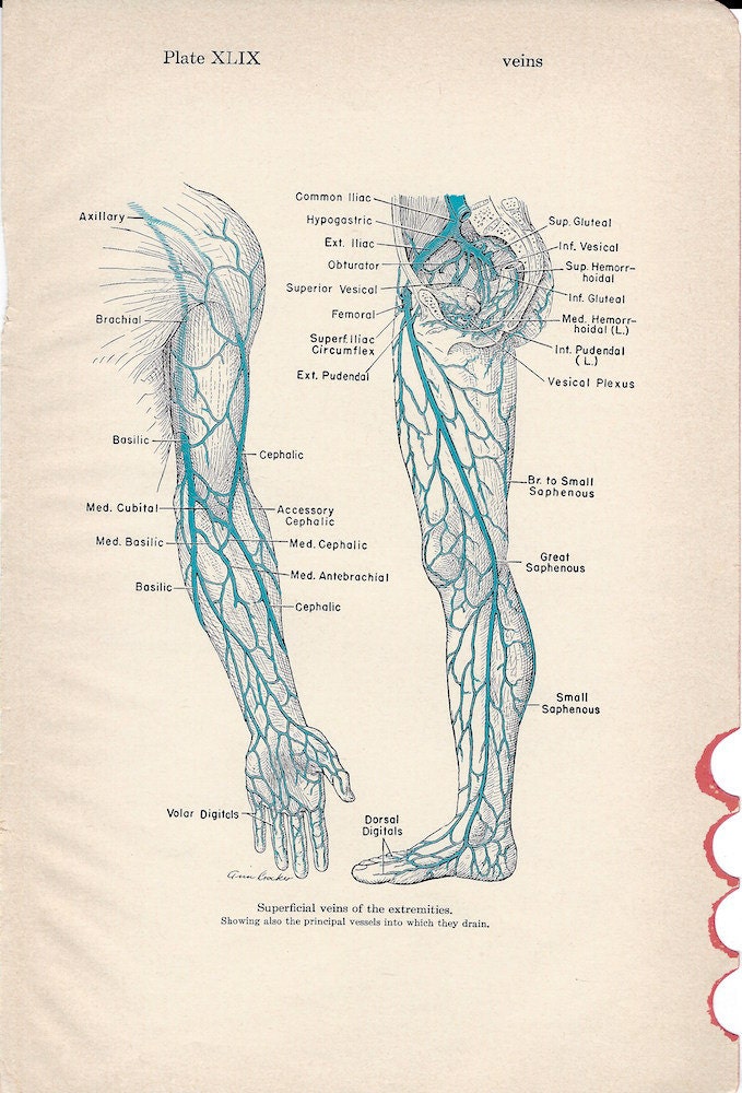
BlogHuman Venous System Anatomy Biology Diagrams Superficial veins, located in the fatty layer under the skin. Deep veins, located in the muscles and along the bones. Connecting veins, which are short veins that link the superficial and deep veins. The deep veins play a significant role in pushing blood toward the heart. The one-way valves in deep veins prevent blood from flowing backward. Structure of Veins. Veins are thin-walled valves containing blood vessels. Anatomically, veins have the same three layered walls as arteries that are the tunica externa, the tunica media, and the tunica intima; however, they have considerably lesser amounts of smooth muscles making them thinner than the walls of the arteries. This thinner wall makes the veins more flexible and allows the veins Explore the venous system with an interactive diagram and learn some tips for improving the health of your veins. Your venous system is a network of veins that carry blood back to your heart from
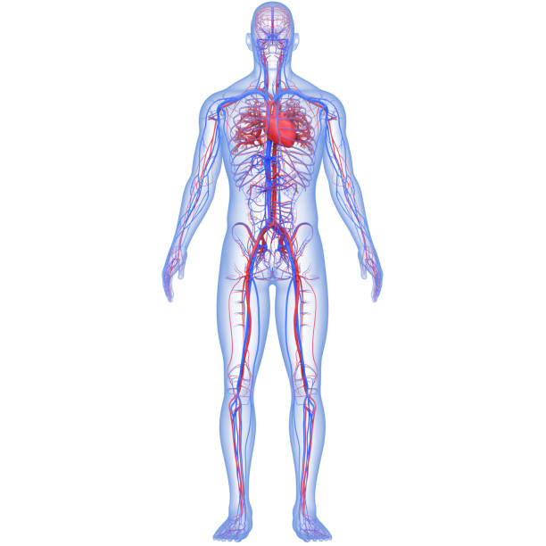
Together, your veins and other blood vessels form a major part of your circulatory system. Your veins connect with venules and capillaries in many places. When mapped out in a drawing, your upper body circulatory system resembles the complex wires and circuits inside a computer. Your lower body circulatory system resembles an upside-down tree The venous system represents a complex network of blood vessels responsible for returning deoxygenated blood from tissues back to the heart. This intricate system comprises numerous interconnected veins varying in size and function, from tiny venules to major vessels like the vena cava. Understanding the venous anatomy is crucial for medical professionals, as it plays a vital role in diagnosis
Physiopedia Biology Diagrams
Veins (/ v eɪ n /) are blood vessels in the circulatory system of humans and most other animals that carry blood towards the heart.Most veins carry deoxygenated blood from the tissues back to the heart; exceptions are those of the pulmonary and fetal circulations which carry oxygenated blood to the heart. In the systemic circulation, arteries carry oxygenated blood away from the heart, and These thin, tube-like structures are an essential part of the circulatory system, which distributes blood and nutrients throughout the body. At any point in time, nearly three-fourths of the circulating blood volume is contained in the venous system. Three types of blood vessels make up the human circulatory system: arteries, veins, and It then flows into a system of capillaries where its exchange functions take place. Figure 15.3.1.2 Human circulation system. Blood from the capillaries flows into venules which are drained by veins. Veins draining the upper portion of the body lead to the superior vena cava. Veins draining the lower part of the body lead to the inferior vena cava.

By studying the venous system diagram and staying up-to-date with advancements in the field, healthcare professionals can provide optimal care and improve patient outcomes. Anatomy of the Venous System. The venous system is an essential part of the circulatory system, responsible for the return of deoxygenated blood back to the heart.
